DicoFlow PACS · Imaging
A diagnostic workstation in every browser
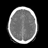
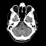
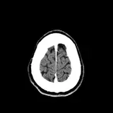
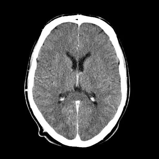
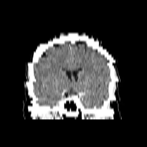
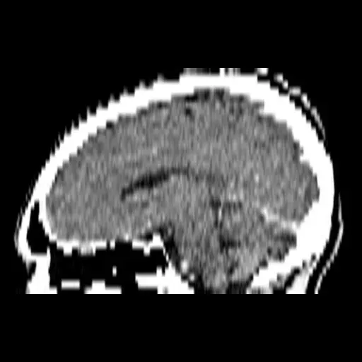
A zero-install DICOM viewer built on Cornerstone3D and VTK.js. Read 2D, MPR and GPU 3D in Chrome, Edge or Firefox on any workstation on your LAN — nothing to install on the reading PCs.
- Cornerstone3D + VTK.js
- WebGL rendering
- Chrome · Edge · Firefox
- Runs on your LAN, offline
By the numbers
One viewer for every study your modalities send
DicoFlow PACS receives, stores and displays images on your own server. Every figure below is counted from DicoFlow LRIMS v2.0.
- DICOM storage SOP classes
- 120
- Interactive viewer tools
- 43
- Window / level presets
- 60
- GPU volume-rendering presets
- 30
Every standard class accepted, × 45 transfer syntaxes
10 viewport layouts, 1×1 to 4×4
Organised in 13 anatomy groups
Plus Smart Auto, filtered to anatomy
2D reading & measurement
The 2D reading tools radiologists expect
Stack-scroll or cine through a series, switch window presets in one click and compare against priors with synchronised viewports. Ten layouts, from 1×1 to 4×4, cover single reads and large comparisons.
- 60 W/L presets in 13 anatomy groups; cine at 1–30 fps
- Key images, sync modes and up to 10 priors side by side
- DICOM tag browser with PHI fields highlighted
- Presenter mode masks patient identifiers on screen
- Touch gestures and a responsive layout
Measurement toolkit
Measure once, keep it with the study
Calibrated measurements, a measurement table and saved annotation sets.
Measurements
Regions of interest
16 specialty measurement services
Measurement calibration
Measurement table
Saved annotation sets
3D, MPR & segmentation
Reformat, render and segment in the same tab
Linked-crosshair MPR, thick-slab projections and GPU volume rendering run in WebGL on the workstation. A capability probe checks each PC first and falls back safely; thin clients can use server-side rendering.
MPR & reformats
- Linked-crosshair MPR
- Thick slab MIP, MinIP or average, 1–50 mm, with 8 presets
- Curved planar reformat from two clicks — the centreline is found automatically
Volume rendering
- GPU rendering with 30 presets, or Smart Auto filtered to the anatomy
- 10 rendering modes
- Bone removal, clipping and VR cut
- Virtual endoscopy
Segmentation
- Brush, eraser, region grow, HU threshold, scissors
- Livewire contour and slice interpolation
- Multi-label, with 3D mesh preview
- Volumes reported in mm³
Export & thin clients
- Isosurface with STL export
- WebM movie export
- Batch sweep saved as a DICOM series
- Server-side rendering for thin clients; GPU probe with safe fallback
Quantitative decision support
Scores, volumes and grades, calculated for you
Rule-based engines do the arithmetic that slows a read down — calcium scores, stroke scores, fracture grades, functional maps. They are decision support, not for primary diagnosis.
Cardiac
Calcium, coronaries and function
Agatston calcium score
Sample- LM
- 0
- LAD
- 112
- LCx
- 18
- RCA
- 46
- Agatston + MESACalcium score per artery, with MESA risk.
- Coronary workspaceCoronary review with CAD-RADS.
- FunctionEjection fraction, stroke volume, cardiac output and LV mass.
- StenosisGraded by the NASCET and ECST methods.
Neuro suite
Ten neuro modules
- 01HaemorrhageICH score · ABC/2
- 02ASPECTS
- 03Brain volumetry
- 04Circle of Willis
- 05AneurysmPHASES
- 06Carotid
- 07Fazekas
- 08PerfusionDAWN · DEFUSE-3
- 09fMRI
- 10Cranial nerves
Spine & bone
Labels you confirm
Automated spine labelling is rule-based and waits for the radiologist to confirm. Vertebral fractures are graded on the Genant scale, and QCT measures bone density.
Chest
Lung nodule detection
Rule-based detection marks candidate nodules on chest CT for the radiologist to review.
Perfusion & diffusion
Functional maps
Quantitative maps open in the same viewer as the source series.
Decision support, not for primary diagnosis. Every engine is rule-based. The radiologist reads the study; the engines do the arithmetic. Engines available by edition — cardiac and neuro on PACS; abdomen, vessel and lung as well on RIMS and LRIMS — licensed as the advanced imaging suite.
Compare editionsHanging protocols & workflow
Studies open ready to read
Series are auto-classified and laid out the moment a study opens. Eight built-in hanging protocols cover routine reads, the Smart Launcher suggests the ones that fit the open study, and the report editor sits beside the images.
- Report editor embedded side-by-side with the viewer
- Saved viewer states are HMAC-signed, so an altered state is detected
- Role-based toolsets — technicians get a focused QC subset
DICOM network & archive
Modalities send. DicoFlow receives, verifies and stores.
A DICOM C-STORE SCP accepts all 120 standard storage SOP classes across 45 transfer syntaxes. Studies are matched to the right patient, encrypted at rest and written to an archive you can verify.
Modalities
…and other DICOM storage senders
C-STORE
Worklist
DicoFlow server · on your LAN
DicoFlow PACS
Windows server PC · works offline
C-STORE SCP + C-ECHO
All 120 standard storage SOP classes × 45 transfer syntaxesListeners & access
Several listeners, each with its own AE title and port. AE whitelist; optional TLS 1.2+.DICOFLOW :104DF_CT :11112Modality Worklist SCP
Patient and order details served to scanners over C‑FINDPatient auto-matching
Unmatched studies wait in a queue for reviewEncrypted at rest
AES-256-GCM, on by default
View/store
Forward
Destinations
Reading workstations
Chrome, Edge or Firefox over the LAN
Local + NAS archive
SHA-256 read-back verified · capacity alerts
Another PACS
C-STORE SCU forwarding
Vendor-neutral export
DICOM ZIP + manifest + conformance statement
Compression with a lossless lock
Optional JPEG 2000 and JPEG-LS rules save disk space. Mammography is locked to lossless.
Capacity you can plan for
Local and NAS tiers with capacity alerts and a growth projection, so you see a full disk coming.
Your data, portable
Vendor-neutral export as a DICOM ZIP with a manifest and conformance statement.
Film studio & patient booklets
Films and booklets, straight from the study
Lay out films in the browser and print to the Windows printers your centre already uses. A safety interlock blocks any film that is missing the patient's name, ID or L/R markers.
Film studio
- 12 film layouts, plus free cell splitting
- 15 film and paper sizes plus custom — 8×10″ to 14×17″, 18×24 to 35×43 cm, A4, A3, Letter, Legal
- Per-image W/L, zoom and rotate; multi-page films; burn-in overlays and a privacy mode
- Printing to Windows / system printers, auto-detected, with live status
- GSDF calibration and a print queue that retries failed jobs
- Print automatically when images arrive or when the study is finalized
Patient booklets
A PDF booklet is generated automatically when the report is verified — branded cover, the signed report with a Code128 barcode, image pages and a back page. Lay it out once in the template designer.
PACS questions
What radiologists and IT leads ask
Short answers on installation, DICOM connectivity, printing and storage.
Read your next study in a browser tab
Book a guided demo of the viewer, the archive and the film studio — on the kind of workstations your radiologists already use.